
Figure 3.
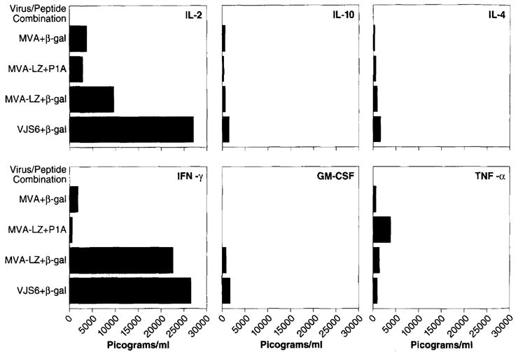
Cytokine profile of splenocytes cultured with MHC class I restricted peptides. Splenocytes were prepared from mice that had previously been inoculated i.m. with 107 p.f.u. of the MVA or MVA-LZ viruses. The same dose of VJS6 given i.v. was used as a positive control. Three weeks later, splenocytes were removed and cultured together with 10 μg ml−1 of the class I-restricted β-gal peptide, TPHPARIGL. Two days later, supernatants of the samples were collected and analyzed by ELISA for IL-2, IFN-γ, IL-10, GM-CSF, IL-4, and TNF-α. Results are shown in picograms per ml of supernatant. Note that each flask contained 30 ml of supernatant