
Fig. 4.
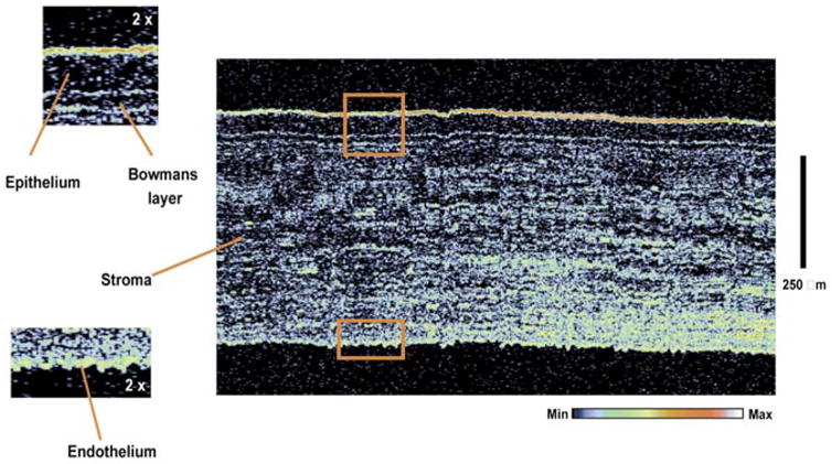
In vivo ultrahigh-resolution corneal OCT image of a normal human subject. The image has 6 × 2 μm (transverse × axial) resolution. The image size is 1.1 × 0.8 mm (transverse × axial) with 600 × 800 pixels, corresponding to 1.8 × 1 μm pixel spacing. The corneal epithelium, Bowman’s layer, intrastromal morphology and endothelium can be visualized. Boxes show enlargement by a factor of two and correspond to matching shapes in large figure.