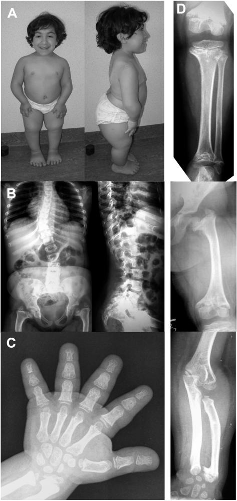
Figure 1. .
Clinical and radiographic features of the novel patient with the AD phenotype. A, Female patient at age 10 years, showing severe disproportionate short stature. B and D, Radiographs of the axial and appendicular skeleton at age 8 years. The spine is abnormal, with thoracolumbar scoliosis and foreshortened vertebral bodies (on lateral film) showing anterior and posterior scalloping at the lumbar level. The end plates are slightly convex. The pelvis shows coxa vara with dysplastic femoral heads and severe shortening of the femoral necks. The femur, tibia, and fibula are short, with severe metaphyseal changes. The knee epiphyses are small and flattened. There is mesomelic shortening in the upper limbs. C, Hand radiograph taken at age 9 years. Metaphyseal changes are apparent in the wrists, with shortening of the distal ulna and ulnar kinking of the distal radius. The phalanges and metacarpals are short and broad (bullet-shaped middle phalanges), with small epiphyses already attached to the metaphyses.

