
Fig. 4. Inactivation of Pax6 inhibits the proliferation of RSCs in vitro.
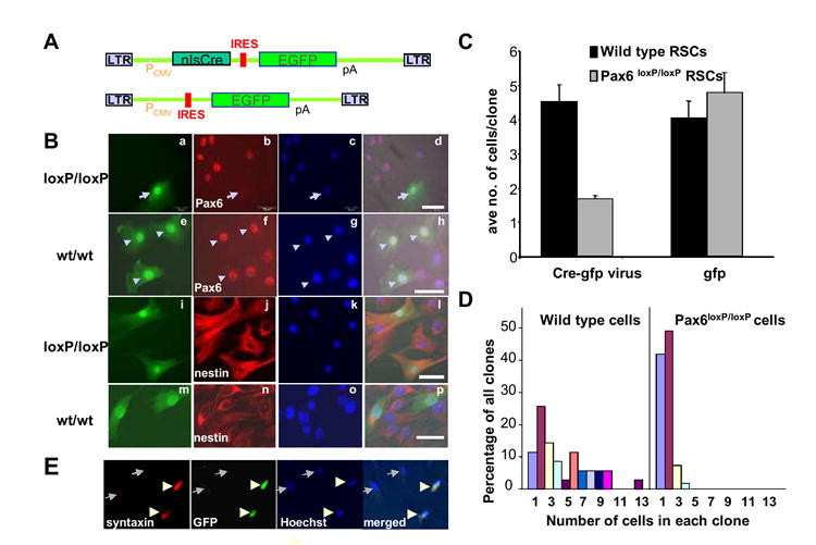
A. pMXIE-Cre-EGFP (top) and pMXIE-EGFP (bottom) retroviral constructs. PCMV: CMV promoter; nlsCre: Cre recombinase with a nuclear localization signal (nls); EGFP: enhanced green fluorescent protein; IRES: internal ribosomal entry site; pA: polyA signal. B. Cre-EGFP retroviral infection of adult Pax6loxP/loxP and wild type RSCs. a-h: Pax6 is inactivated in the Cre-EGFP infected Pax6loxP/loxP RSCs (arrows in a-d), but not the wild type RSCs (arrowheads in e-h). a,e: GFP channel showing the infected cells; b,f: Pax6 immunostaining; c,g: Hoechst nuclear staining; d,h: merged images. i-p: RSCs infected with Cre-EGFP retrovirus retain progenitor identity with neural progenitor marker nestin expression. i,m: GFP channel showing the infected cells; j,n: immunostaining of nestin. k,o: Hoechst nuclear staining; l,p: merged images. GFP expression marks the infected RSCs and their progeny. C: Average number of progeny in each clone derived from single viral infected adult RSCs 4 days after retroviral infection in vitro. Data represent means +/- SEMs. D. Distribution of different sized clones derived from single virus-infected RSCs 4 days after Cre-EGFP retroviral infection of wild type or Pax6loxP/loxP ciliary epithelial cells. E. Immunohistochemistry of differentiated RSCs from Pax6loxP/loxP animals 21 day after pMXIE-Cre-EGFP viral infection with syntaxin monoclonal antibody. Arrowheads showed two infected cells, which are expressing the amacrine cell marker syntaxin. Arrows pointed two non-infected cells, which were syntaxin negative.