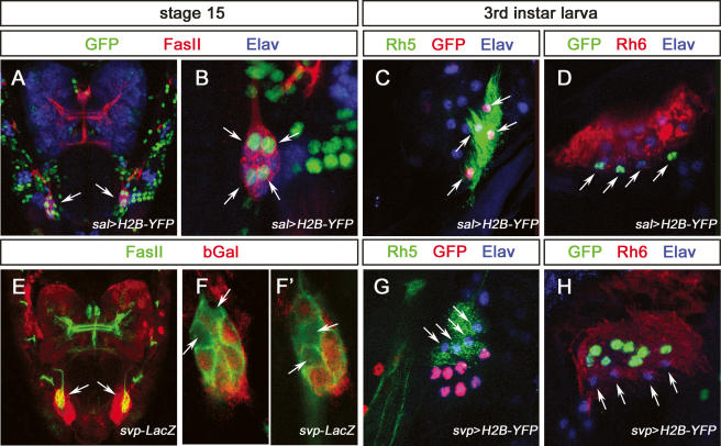
Figure 5.
Expression of sal and svp in the developing and mature larval PRs. (A) Dorsal view of a sal-Gal4,UAS-H2B-YFP stage 15 embryonic head labeled with anti-GFP (green), anti-FasII (red), and anti-Elav (blue), and GFP expression in BO (arrow). (B) High-magnification image of the BO in A. Four cells are labeled by anti-GFP staining (arrow). (C) High-magnification image of BO in third instar larva sal-Gal4,UAS-H2B-YFP, labeled with anti-GFP (red), anti-Rh5 (green), and anti-Elav (blue). Anti-GFP labeling coincides with anti-Rh5 staining (arrow). (D) High-magnification image of BO in third instar larva sal-Gal4,UAS-H2B-YFP, labeled with anti-GFP (green), anti-Rh6 (red), and anti-Elav (blue). Anti-GFP labeling is excluded from anti-Rh6 staining (arrows). (E) Dorsal view of a svp-lacZ stage 15 embryonic head labeled with anti-FasII (green) and anti-βGal (red) expression in BO (arrows). (F,F′) High-magnification image of the BO in E; individual optical sections show four cells are devoid of anti-βGal staining (arrows). (G) High-magnification image of BO in third instar larva svp-Gal4,UAS-H2B-YFP, labeled with anti-GFP (red), anti-Rh5 (green), and anti-Elav (blue). Anti-GFP labeling is excluded from anti-Rh5 staining (arrows). (H) High-magnification image of BO in third instar larva svp-Gal4,UAS-H2B-YFP, labeled with anti-GFP (green), anti-Rh6 (red), and anti-Elav (blue). Anti-GFP labeling coincides with anti-Rh6 staining, and is excluded for the remaining four PRs (arrows).

