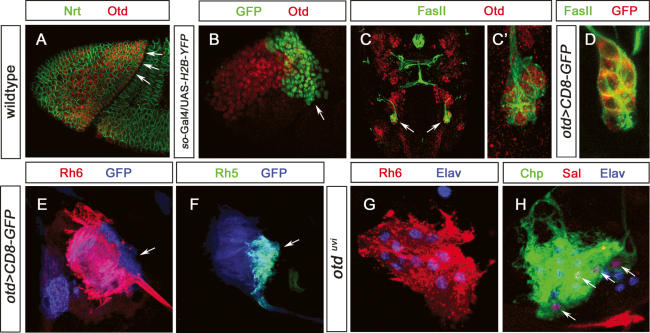
Figure 7.
Expression and function of otd in developing and mature larval PRs. (A) Lateral view of a stage 7 embryo (procephalic region) stained with anti-Nrt (green) and anti-Otd (red); the region giving rise to the optic lobe anlage and larval PRs expresses Otd (arrow). (B) Lateral view of a so-Gal4/UAS-H2B-YFP stage 10 embryonic head region stained with anti-GFP (green) and anti-Otd (red); the region giving rise to larval PRS (ventral/lateral tip) is devoid of Otd expression. (C) Dorsal view of a wild-type stage 15 embryonic head labeled with anti-FasII (green), anti-Otd (red), and anti-Otd staining in BO, indicated by arrows. (C′) High-magnification image of C. All immature PRs are labeled by anti-Otd staining. (D) High-magnification image of embryonic stage 15 otd-Gal4/UAS-CD8∷GFP BO labeled with anti-FasII (green) and anti-GFP (red). All immature PRs are labeled by anti-GFP staining. (E) High-magnification image of third instar larva otd-Gal4/UAS-CD8∷GFP BO labeled with anti-GFP (blue) and anti-Rh6 (red). (F) High-magnification image of third instar larva otd-Gal4/UAS-CD8∷GFP BO labeled with anti-GFP (blue) and anti-Rh5 (green). (G) High-magnification image of third instar larva otdvui mutant BO labeled with anti-Rh6 (red) and anti-Elav (blue). All PRs are labeled by anti-Rh6 staining. (H) High-magnification image of third instar larva otdvui mutant BO labeled with anti-Chp (green), anti-Sal (red), and anti-Elav (blue). Four PRs are labeled by anti-Sal staining.

