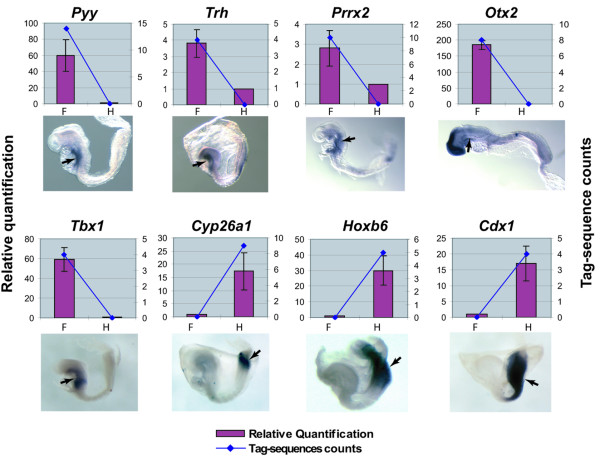
Figure 3.
Correlation of the expression validation of 8 genes from the first list between RT-qPCR, whole mount in situ hybridization and SAGE. For each gene, the upper panel shows the comparison of expression level using RT-qPCR and SAGE (Left scale: relative quantification indicated by the bars; Right scale: raw tag-sequence counts indicated by the line. F: foregut; H: hindgut). The lower panel shows the expression pattern detected by whole mount in situ hybridization. For all embryos, anterior is to the left and posterior is to the right. The RT-qPCR, whole mount in situ hybridization and SAGE validation results were well correlated. pYY, Trh, Prrx2, Otx2 and Tbx1 are highly expressed in the foregut (indicated by arrow). Conversely, Cyp26a1, Hoxb6 and Cdx1 are highly expressed in the hindgut (indicated by arrow).

