
Figure 2.
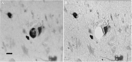
Nestin-labeled giant cell (arrow) visualized before (A) and after (B) microdissection and aspiration. Note adjacent neuron unaffected by procedure. In A, section is viewed under water, while in B the section has been dehydrated and coverslipped. (Bar = 40 μm.)