
Figure 4.
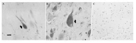
(A and B) Neurons (A) and giant cells (B) labeled with nestin antibodies (arrows). Neuronal staining was within the somatodendritic region and proximal axonal segment. (Bar = 30 μm.) (C) Absence of nestin staining in control temporal cortex.