Figure 4.
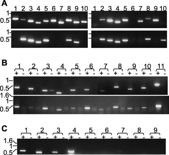
Analysis of VR genes. (A) Presence of VR sequences in several T. brucei strains. PCR analysis of genomic DNA was undertaken with primers specific for: lanes 1–10, VR 1, 2, 4, 5, 8, 9, 11, 13, 15, and 18. T. brucei genomic DNA sources: top left, TREU 927; top right, Lister 427; bottom left, STIB 247; bottom right, EATRO 795. (B) Lack of stage-specificity of VR transcripts. RT-PCR analysis of VR transcripts represented in oligo[dT]-primed cDNA from bloodstream stage (top) and procyclic stage (bottom) TREU 927. Lane pairs 1–10: primers for VR 1, 2, 4, 5, 8, 9, 11, 13, 15, and 18; lane pairs 11: tubulin primers. In each lane pair, “+” denotes presence, and “−” denotes absence, of reverse transcriptase at the cDNA synthesis step. (C) Analysis of whether transcripts of alleles of VR and VSG genes are mutually exclusive. RT-PCR analysis of Lister 427 bloodstream trypanosome clone expressing linked HYG and 221 VSG genes, grown in hygromycin. oligo[dT]-primed cDNA was subjected to PCR using (lane pairs 1–9) primers for VR 2, VR 5, VR 15, 221 VSG, 118 VSG, VO2 VSG, 121 VSG, G4 VSG, S8 VSG. All images are of ethidium bromide-stained gels, and DNA size markers (kb) are indicated to the left of each.

