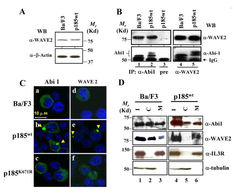
Fig. 2.

Complex formation and membrane translocation of Abi1/WAVE2 in Ba/F3 cells transformed by p185wt. A. Expression of WAVE2 in Ba/F3 and p185wt-transformed Ba/F3 cells. Total lysates from 2×10⁵ cells were analyzed by Western blot using indicated antibodies. B. Complex formation of Abi1 and WAVE2. The lysates from Ba/F3 cells transduced with either an empty retroviral vector (control) or the retroviral vector expressing p185wt were immunoprecipitated with anti-Abi1 antibody, anti-WAVE2 antibody, or pre-immune rabbit serum (pre), as indicated. The immunoprecipitates were analyzed by Western blot using indicated antibodies. C. Bcr-Abl-induced membrane translocation of Abi1/WAVE2. Retroviral vectors expressing GFP-Abi1 (panels a-c) and GFP-WAVE2 (panels d-f) were introduced into Ba/F3 cells or Ba/F3 cells expressing wild type and the mutant form of p185Bcr-Abl, as indicated. The subcellular localization of GFP-Abi1 and GFP-WAVE2 was analyzed by two-photon confocal microscopy. Nuclei were stained by DAPI (blue). Arrowheads indicate the membrane localization of Abi1 and WAVE2. Bar: 10 μm. D. Subcellular distributions of Abi1 and WAVE2 in Ba/F3 and the Ba/F3 transformed by p185wt. The Ba/F3 cells and the Ba/F3 cells transformed by p185wt were lysed and fractionated to separate the cytosol (C) and plasma membrane (M). The equal amounts of total lysate (I, input), cytosol, and membrane fractions were separated on SDS-PAGE and analyzed by Western blot using antibodies specific to Abi1 and WAVE2, as indicated. To monitor the quality of the fractionation, the blot was also probed with the antibodies to the β subunit of IL-3 receptor and α-tubulin, the proteins known to be in membrane and cytosol, respectively.
