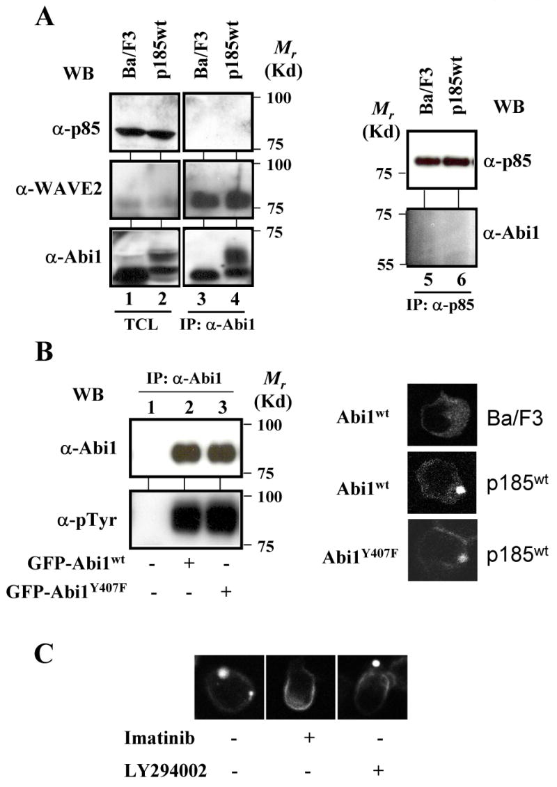
Fig. 5.

The PI3K pathway is not required for Bcr-Abl-induced membrane translocation of Abi1. A. Abi1 does not form complex with p85 subunit of PI3K in Ba/F3 cells and the Ba/F3 cells transformed by p185wt. The lysates from 2×10⁷ Ba/F3 cells and the Ba/F3 cells transformed by p185wt were immunoprecipitated by anti-Abi1 (lanes 3 and 4, left panel) and anti-p85 (lanes 5 and 6, right panel) antibodies, respectively. The immunoprecipitates were analyzed by Western blotting using the indicated antibodies. A portion of total cell lysates (TCL) equivalent to 2×10⁵ cells (p85 and WAVE2), or 1×10⁶ cells (Abi1) was also analyzed by Western blotting to show the expression level of Abi1, p85, and WAVE2 (lanes 1 and 2, left panel). B. The mutation at tyrosine 407 does not affect the Bcr-Abl-induced membrane translocation of Abi1Y407F. Left panel: lysates from p185wt-transformed Ba/F3 cells (lane 1) and the p185wt-transformed Ba/F3 cells expressing either GFP-Abi1 (lane 2) or GFP-Abi1Y407F (lane 3) were immunoprecipitated by anti-Abi1 antibody and analyzed by Western blotting using the antibodies indicated. Right panel: Ba/F3 cells and Ba/F3 cells transformed by p185wt, as indicated, were transduced with retroviruses expressing either GFP-Abi1 or GFP-Abi1Y407F. The subcellular distribution of GFP-fusion proteins was visualized by 2-photon confocal microscopy. C. LY294002 failed to inhibit Bcr-Abl-induced membrane translocation of GFP-Abi1. The p185wt-transformed Ba/F3 cells were transduced with the retrovirus expressing GFP-Abi1. The cells were then left untreated or treated with either 10 μM imatinib or 50 μM LY294002, as indicated, for 8 hours. The cells were fixed and counterstained with TRITC-conjugated phalloidin and DAPI to visualize the F-actin and nuclei, respectively. Images were captured by two-photon confocal microscopy. A color picture is presented in supplemental Fig. 2B.
