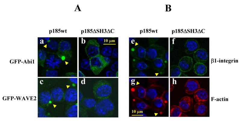
Fig. 6.

The p185ΔSH3ΔC failed to induce the membrane translocation of Abi1/WAVE2, integrin clustering, and abnormal actin remodeling. A. Ba/F3 cells expressing either p185wt (a and c) or p185ΔSH3Δc (b and d) were transduced with the retroviral vectors expressing either GFP-Abi1 (a and b) or GFP-WAVE2 (c and d). The cells were fixed and GFP-fusion proteins were visualized by 2-photon confocal microscopy. B. Ba/F3 cells transduced with either p185wt (e and g) or p185ΔSH3ΔC (f and h) were fixed and incubated with FITC-conjugated monoclonal antibody specific for β1-integrin (e and f). The cells were also stained with TRITC-conjugated phalloidin to visualize F-actin (g and h). The nuclei of the cells shown in C and D were stained by DAPI (blue) and the arrowheads indicate the subcellular localization of GFP-Abi1 (a), WAVE2 (c), clustering β1-integrin (e), as well as abnormal actin-enriched structures (g) in p185wt-transformed cells. Bar: 10 μm.
