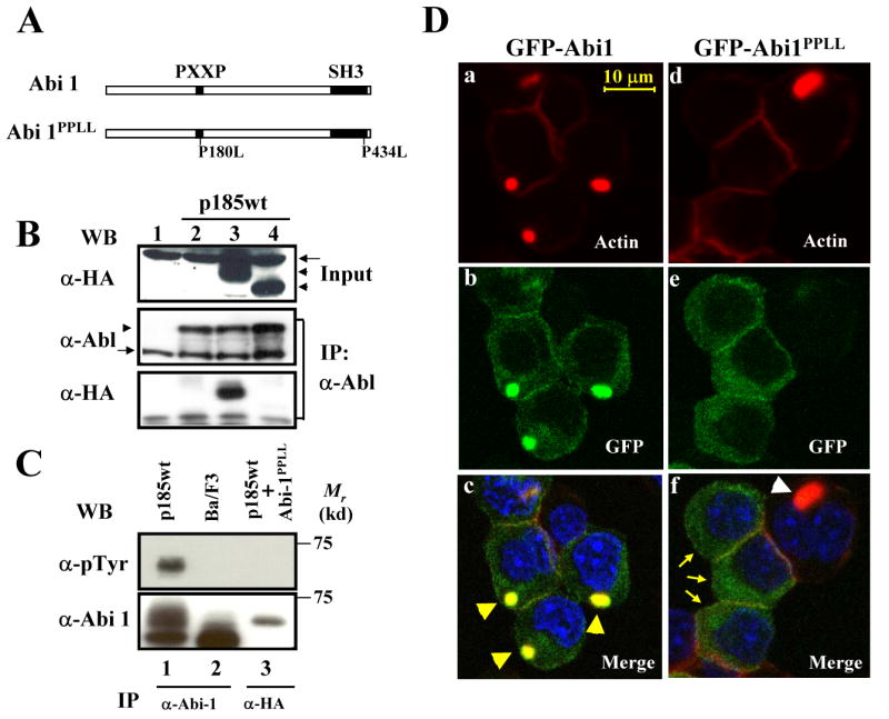
Fig. 7.

Expression of Abi1PPLL inhibited Bcr-Abl-induced abnormal actin cytoskeleton remodeling. A. Schematic diagram of Abi1 and Abi1PPLL. B. Abi1PPLL is defective in binding to Bcr-Abl. Ba/F3 cells (lane 1) and the Ba/F3 cells expressing p185wt alone (lane 2), p185wt plus HA-tagged Abi1 (lane 3), and p185wt plus HA-tagged Abi1PPLL (lane 4) were lysed and immunoprecipitated with anti-Abl antibody. The immunoprecipitates (middle and lower panels) and 1/50 of total cell lysates used for IP (Input, upper panel) were analyzed by Western blot using indicated antibodies. The HA-tagged Abi1 and Abi1PPLL are indicated by arrowheads, whereas a non-specific band cross-reacted with anti-HA antibody is indicated by arrow (upper panel). The anti-Abl antibody recognized both Bcr-Abl and endogenous c-Abl, as indicated by arrowhead and arrow, respectively (middle panel). C. Abi1PPLL failed to be tyrosine-phosphorylated by Bcr-Abl. Ba/F3 cells (lane 2) and Ba/F3 cells expressing p185wt alone (lane 1) or p185wt plus HA-tagged Abi1PPLL (lane 3), were lysed and immunoprecipitated with indicated antibodies. The immunoprecipitates were analyzed by Western blot using indicated antibodies. D. Inhibition of Bcr-Abl-induced abnormal actin remodeling by Abi1PPLL. Ba/F3 cells transformed by p185wt were transduced with retroviruses expressing either GFP-Abi1 (a-c) or GFP-Abi1PPLL (d-f). Cells were fixed and stained with TRITC-conjugated phalloidin and DAPI to visualize F-actin (red) and nuclei (blue), respectively. Subcellular localization of GFP-fusion proteins (b, e, green) and F-actin structure (a, d, red) were analyzed by two-photon confocal microscopy. The merged images were also shown (c, f). The arrowheads in panel c indicate abnormal actin-enriched structures that co-localize with GFP-Abi1, whereas arrows in panel f indicate the p185wt-transformed cells that express GFP-Abi1PPLL. An open arrowhead in panel f indicates abnormal actin-enriched structure in a cell that did not express Abi1PPLL. Bar: 10 μm.
