Figure 4.

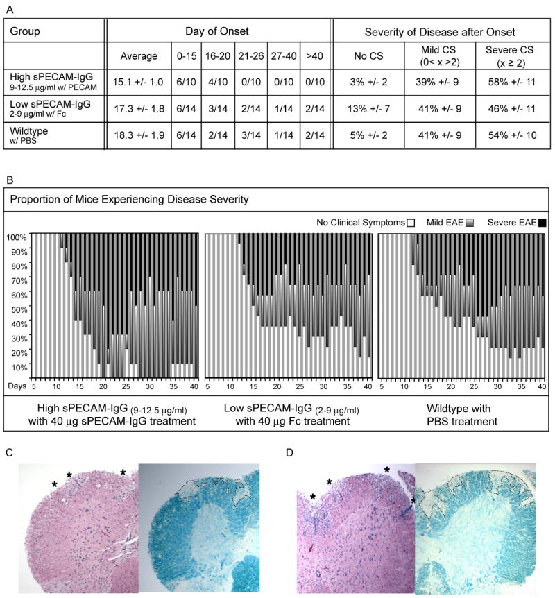
sPECAM-Fc treatment of high sPECAM-Fc expressing mice increases disease onset but not disease severity as compared to wildtype mice. High sPECAM-Fc expressors, low sPECAM-Fc expressors, and wildtype mice were treated 5,7,9,11,13 days after induction of EAE. High expressors received 40μg of purified sPECAM-Fc; low expressors received 40μg of human IgG-Fc; wildtype mice received PBS alone. A. Shown is average day onset, plus or minus standard error of the mean, and the number of mice per group to experience onset within the given time frames for all three groups. For wildtype to high comparison, p= 0.06, for all other comparisons, p > 0.1 The average percentage of time during the fifteen days after onset that mice in each group experienced no clinical symptoms, mild EAE with clinical symptoms greater than zero and less than 2, and severe EAE with clinical symptoms of 2 and above is shown. B. Graphical representation shows the proportion of mice in the group on each day experiencing no onset or no clinical symptoms (white portion of bar), mild EAE (red portion of bar), or severe EAE (black portion of bar). (C) Low sPECAM-Fc expressor and (D) high sPECAM-Fc expressor. Spinal cords were removed 40 days post EAE induction. Paraffin-embedded sections were stained with H&E or LFB to detect infiltrating mononuclear cells and demyelinated areas, respectively. Both low and high expressors showed visible signs of infiltration and demyelination in the spinal cord.
