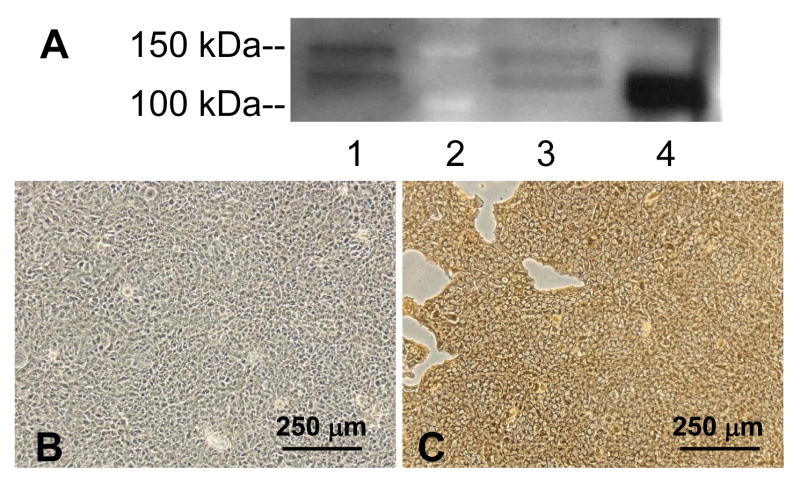
Figure 3.
APP protein is expressed in the choroid plexus tissue and the choroidal epithelial cell line. A) Representative Western blot for APP presence in Z310 cells (lane 1), choroid plexus (lane 3) and homogenized whole brain (lane 4; positive control). Lane 2 is the molecular weight markers for 100 and 150 kDa. Bands are present at 110 kDa. Results are not quantitative. B) Negative control in which the primary antibody against APP was excluded from the immunocytochemistry in Z310 cells. C) APP expression (brown stain) in Z310 cells. B and C are representative of three immunocytochemistry experiments.

