
Figure 5.
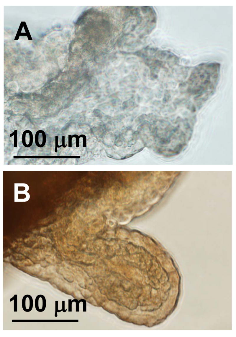
Immunohistochemistry of neprilysin in the choroid plexus. Neprilysin expression is evident in choroid plexus tissue (B) compared to a control tissue, in which the primary antibody was excluded (A). These images are representative of at least three trials.