
Figure 4. Noggin enhances neural crest migration into the hindgut.
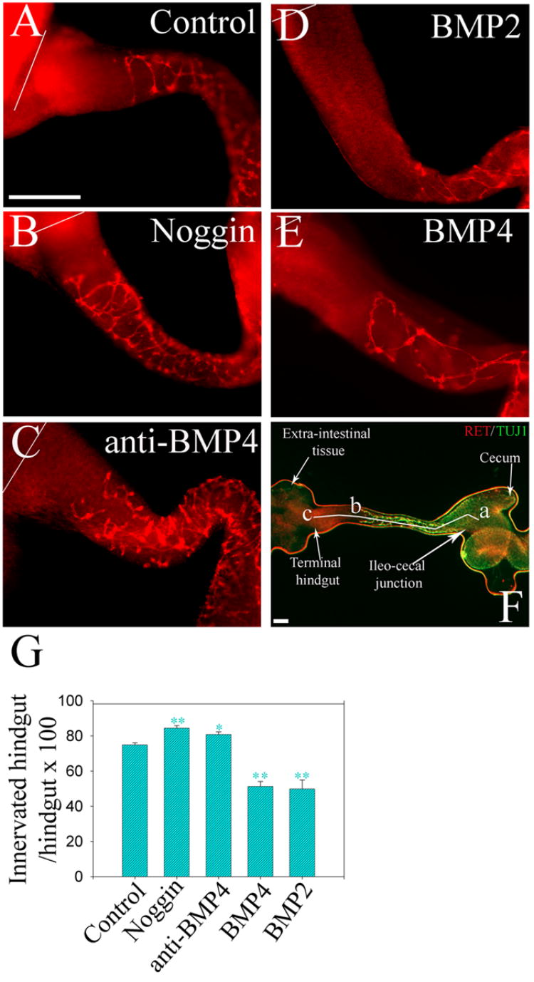
E11.5 mouse gut explants including the esophagus, stomach, small bowel and colon were cultured for 48 hours (A) under control conditions, or (B) with noggin, (C) anti-BMP4 blocking antibody, (D) BMP2, or (E) BMP4. Whole mount immunohistochemistry for Ret demonstrated NCC within the bowel. White lines indicate the end of the colon. (F) Shows landmarks for measurements: segment “a–b” = the region of colon colonized by NCC; segment “a–c” = the whole length of the hindgut. “a” is the ileo-cecal junction; “b” is the position of the most distal Ret expressing cell at the migration wave front. “c” is the terminal hindgut. Also seen is the extra-intestinal tissue we used to secure the explants to the gel bed. (G) Quantitative analysis demonstrated that noggin or anti-BMP4 blocking antibody treatment increased the percentage of the colon innervated, while BMP treatment reduced the extent of colon innervation. Scale bar = 100 μm. * P<0.001; **:P<0.01.