
Figure 5.
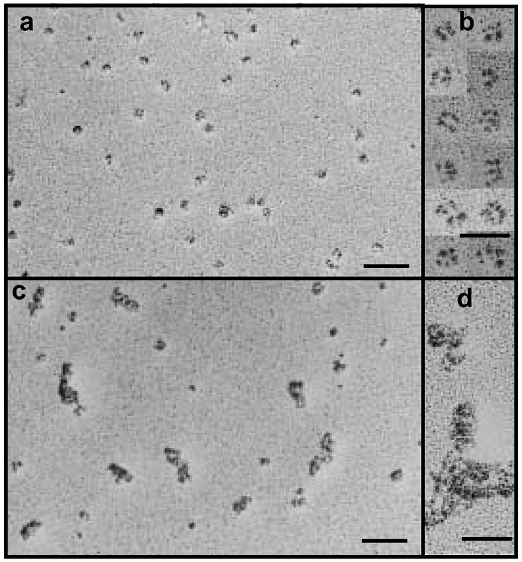
Analysis of the structure of hFcILZOX40L by rotary shadowing and electron microscopy. (a, b) hFcILZOX40L eluted form protein-g at neutral pH; (c, d) hfcILZOX40L eluted from protein-G at acidic pH. The scale bars for the fields in a and c are 100 nm and for the selected images in b and d, 50 nm.