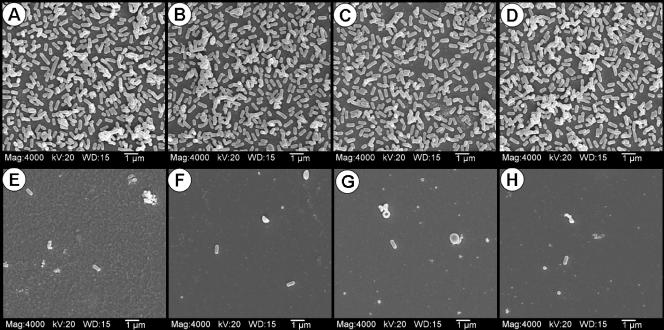
FIG. 3.
SEM images of various strongly and weakly adherent strains of L. monocytogenes screened using the microplate attachment assay with glass chips. The strains of L. monocytogenes are as follows: top row, CW50 (A), CW62 (B), CW77 (C), and 99-38 (D); bottom row, CW34 (E), CW35 (F), CW52 (G), and SM3 (H). Mag, magnification; WD, working distance (mm).

