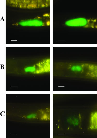
FIG. 3.
Epifluorescent images of the level of colonization (high [A], medium [B], and low [C]) of the distal area of the receptacle in S. carpocapsae IJs. In all panels, the anterior end of the nematode is oriented to the left side of the panel. Left and right panels are images representative of the different levels of colonization. Bars, 10 μm.

