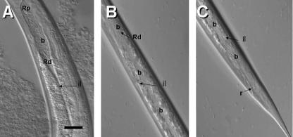
FIG. 6.
DIC images showing bacterial release process. Shown is the opening of the bacterial receptacle into the intestine. Notice the expansion of the intestinal lumen (il) and the migration of the bacterial cells (b) into this lumen. The anterior end of the nematode is oriented to the top of the panel. Bar, 6 μm.

