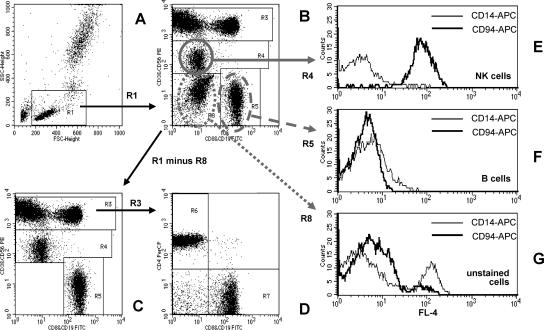
FIG. 1.
Quantification of peripheral blood lymphocyte subsets by the T1 FCM method. (A) Lymphocytes are identified as low FSC and low SSC cells (region R1). Details of the gating strategy are described in the text. (B) Region R8 contains cells that did not stain with antibodies against CD3, CD8, CD56, or CD19. This cell population is excluded from analysis by defining the lymphocyte gate G1 for R1 and R2 (R2 includes R3 and R4 and R5 but not R8). (C and D) The dot plots for the final analysis are shown. (E and F) The purity of gating of NK and B cells is shown. (E) The NK cell gate (R1 and R4) contains mainly CD94+ NK cells and very few CD14+ monocytes. (F) Neither CD94+ NK cells nor CD14+ monocytes are found in the B-cell gate (R1 and R5). (G) Cells that did not stain with antibodies against CD3, CD8, CD56, or CD19 include CD14+ monocytes, as well as CD56− CD94+ NK cells.

