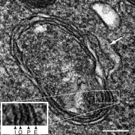
FIG. 1.
Transmission electron micrograph of a T. gondii apicoplast. The inset shows an enlargement of the four membranes visible in the image, marked according to their origin. I, derived from inner chloroplast membrane of cyanobacterial origin; O, derived from outer chloroplast membrane of cyanobacterial origin; P, periplastid membrane of algal plasma membrane origin; and E, outer membrane of apicomplexan endomembrane system origin. The bulge on the side of the outer membrane of the apicoplast is marked with an arrow. Bar = 100 nm.

