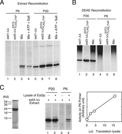
FIG. 4.
Reconstitution of full telomerase activity from mutant extracts in vitro. (A) Extracts derived from an est1Δ/est1Δ (est1-ΔΔ) and an est3Δ/est3Δ (est3-ΔΔ) EST2-FLAG3/TAP strain were either separately subjected to IgG-Sepharose pulldown or subjected to pulldown following mixing. Telomerase activity on the beads was analyzed by primer extension assays using either the P6 or the P20 primer. (B) DEAE fractions (400 mM to 900 mM) were derived from the same strains as those used for panel A. The fractions were assayed separately or after mixing as indicated at the top of the gel. (C) (Left) Labeled Est3p obtained by in vitro transcription and translation was analyzed by sodium dodecyl sulfate-polyacrylamide gel electrophoresis and a PhosphorImager. (Center) Extracts derived from the est3Δ/est3Δ EST2-FLAG3/TAP strain were mixed with 15 μl of mock-translated lysates or Est3p-containing lysates. Telomerases in the mixtures were isolated on IgG-Sepharose and subjected to primer extension using the indicated primers. (Right) Extracts derived from the est3Δ/est3Δ EST2-FLAG3/TAP strain were mixed with increasing amounts of Est3p-containing lysates. Telomerases in the mixtures were isolated on IgG-Sepharose and subjected to primer extension using the P6 primer. The signals were quantified and plotted.

