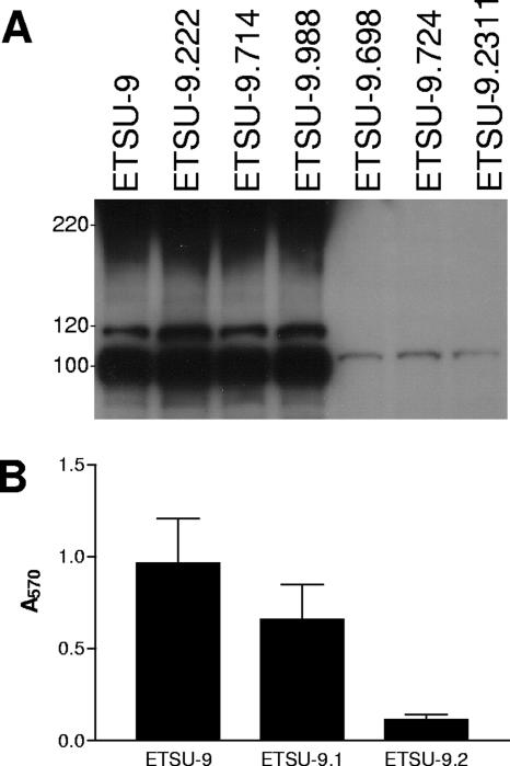
FIG. 1.
Characterization of backcross-derived, biofilm-negative M. catarrhalis ETSU-9 mutants. (A) Detection of UspA2H expression by biofilm-negative ETSU-9 transposon mutants. Whole-cell lysates of the ETSU-9 parent strain and the six biofilm-negative transposon mutants were probed in Western blot analysis for UspA2H expression using MAb 17C7 as the primary antibody probe. The UspA2H protein appears as a smear around the 220-kDa position marker and as several smaller MAb-reactive species with apparent molecular masses of 118 kDa and 100 kDa in the Western blot. The UspA1 protein is visible in the last three lanes as a faint band just above the 100-kDa position marker. Molecular mass position markers (in kDa) are on the left. (B) Biofilm formation by wild-type and mutant strains of ETSU-9. Biofilm formation by the ETSU-9 parent strain, the uspA1 mutant ETSU-9.1, and the uspA2H mutant ETSU-9.2 was measured using the crystal violet-based assay. Each strain was grown in three different wells per experiment, and the data represent the means and standard errors from three independent experiments.

