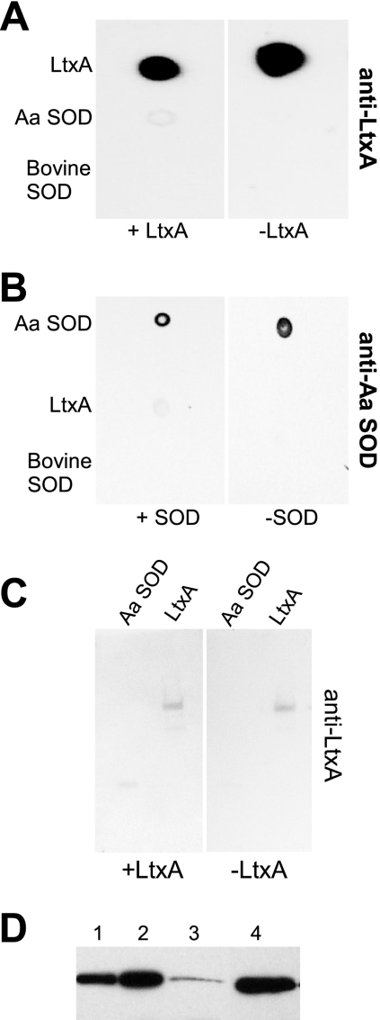
FIG. 3.
Protein interaction assays. (A and B) Dot blot assays. Purified proteins (0.5 μg) were spotted onto a nitrocellulose membrane. The membrane was blocked and then incubated with purified LtxA (50 μg/ml) (A) or Cu,Zn SOD (B). Membranes that were not treated with LtxA (A) or Cu,Zn SOD (B) served as negative controls. The membranes were subjected to Western blot analysis with anti-LtxA (A) or anti-Cu,Zn SOD (B) antibody. (C) Overlay assay. Purified proteins (0.5 μg) were resolved by SDS-PAGE and then transferred to a nitrocellulose membrane. The membrane was incubated with purified LtxA (50 μg/ml). A membrane that was not incubated with LtxA served as a negative control. The membranes were subjected to Western blot analysis with anti-LtxA antibody. (D) Localization of Cu,Zn SOD. Cells were fractionated with the detergent sarkosyl. Lanes: 1, periplasm; 2, cytosol; 3, membrane; 4, purified Cu,Zn SOD (2.5 μg). Aa SOD, A. actinomycetemcomitans Cu,Zn SOD; Bovine SOD, bovine Cu,Zn SOD.

