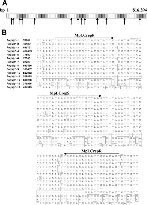
FIG. 1.
(A) Schematic representation showing the location of the different repMp1 copies (indicated by arrows) in the genome of M. pneumoniae strain M129. (B) Partial alignment of the 14 repMp1 copies of M. pneumoniae strain M129 and the location of the primer (drawn arrow) and of the probe (dashed arrow) used for real-time PCR. Differing bases and gaps are indicated by boxes. Genome positions are derived from the published sequence (7; GenBank accession no. U00089).

