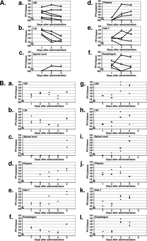
FIG. 5.
Titers of PV recovered in tissues after oral administration of PV. (A) Virus was extracted from tissues of mice (PVRTg21 [open triangles], PVRTg21/IfnarKO [solid triangles], MPVRTg25 [open circles], MPVRTg25/IfnarKO [solid circles], C57BL/6 [open squares], and C57BL/6/IfnarKO [solid squares]) 1, 2, and 3 days after the administration of 3 × 10⁸ PFU of PV1(M)OM/2 ml. The vertical axis shows the amount (PFU) of PV detected in tissues by the plaque assay. USI, upper small intestine; LSI, lower small intestine. (B) Virus was extracted from each tissue of PVRTg21/IfnarKO every 24 h after oral administration of 3 × 10⁸ PFU of PV1(M)OM/2 ml (a to f) or intravenous injection of 1 × 10⁵ PFU of PV1(M)OM/100 μl (g to l). Each point indicates one mouse. ND, not detected.

