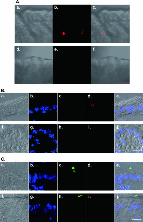
FIG. 6.
Fluorescently labeled PV is detected in intestinal epithelia. Alexa Fluor 555-labeled PV1(M)OM (1.5 × 10⁷ PFU/10 μl) was injected into the ligated small intestines of MPVRTg25/IfnarKO (top row of images in each panel). PV-negative controls were also used (bottom row in each panel). One hour after injection, the ligated portion was excised and subjected to confocal laser scanning microscopy. Intestines were observed without (A) or with (B and C) fixation and frozen sectioning. (B) UEA-1-negative epithelia; (C) epithelia that contain UEA-1-positive cells. Red, Alexa Fluor 555-labeled PV1(M)OM (Ab, c, e, and f; Bd, e, i, and j; Cd, e, i, and j); blue, nuclei (Bb, e, g, and j; Cb, e, g, and j); green, fluorescein isothiocyanate-labeled UEA-1 (Bc, e, h, and j; Cc, e, h, and j). Aa and d, Ba and f, and Ca and f show bright-field images only. Ac and f, Be and j, and Ce and j show merged images of bright-field and fluorescence micrographs. Bars, 100 μm.

