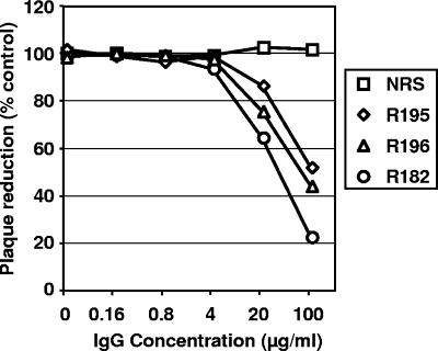
FIG. 3.
Inhibition of plaque formation by anti-B6 PAbs. BSC-1 cell monolayers were infected with EVs that were previously incubated for 1 h with the indicated antibody (along with anti-MV neutralizing antibodies). After 18 h of incubation at 37°C, the cells were fixed and stained with crystal violet, and plaques were counted. Data are expressed as the percentage of plaque reduction relative to the control with no IgG. Each plotted point represents an average of two wells. NRS, normal rabbit serum IgG. R195 and R196 are rabbit PAbs to B6t; PAb R182 is a rabbit PAb to B5. This experiment was done twice with the same results.

