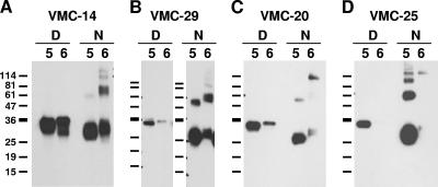
FIG. 5.
Patterns of MAb recognition of B5t and B6t by Western blotting. Purified baculovirus-expressed B5t (indicated by a 5) or B6t (indicated by a 6) was electrophoresed on a 12% Tris-glycine polyacrylamide gel under denaturing (D) or nonreducing (N) conditions, transferred to nitrocellulose and probed with each of the purified MAbs. Representative recognition patterns are shown. (A) Strong recognition of both proteins; (B) strong recognition of B6t under nonreducing conditions; (C) weak recognition of B6t under both conditions; (D) no recognition of B6t. The sizes of molecular mass markers are shown in kDa.

