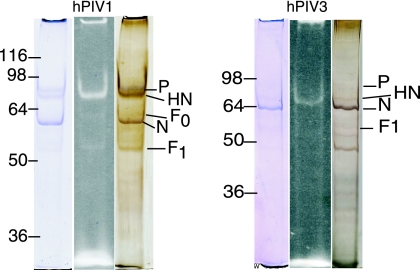
FIG. 1.
Sodium dodecyl sulfate gel electrophoresis of Alexa-labeled hPIV1 and hPIV3. The fluorescent band (center lane) corresponding to HN is compared to lanes stained with Coomassie blue (left) or Bio-Rad silver reagent (right). Numbers to the left of each panel are molecular masses in kilodaltons.

