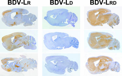
FIG. 4.
Growth characteristics in mouse brains of BDV variants with amino acid changes in L. Virus antigen distribution in sagittal brain sections of infected MRL mice is shown. Three animals per group were infected as newborns with 1,000 FFU of the indicated recombinant viruses and sacrificed 8 weeks later. Virus propagation was analyzed by immunohistochemistry using a rabbit antiserum specific for BDV-N. The brown stain indicates BDV-infected cells.

