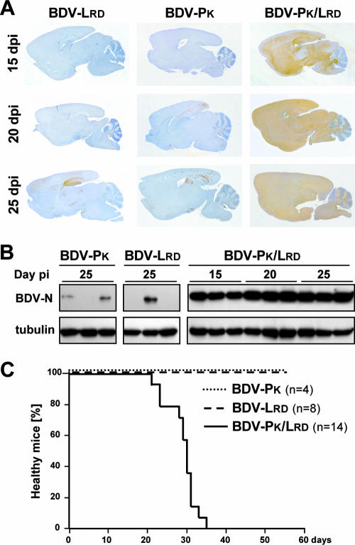
FIG. 5.
P mutation R66K stimulates propagation and pathogenicity of BDV in mice. C57BL/6-IFNAR10/0 mice were infected with 1,000 FFU of the indicated recombinant viruses. At 15, 20, or 25 days postinfection, three animals of each group were sacrificed. (A) Virus propagation in one brain hemisphere was analyzed by immunohistochemistry using a rabbit antiserum specific for BDV-N. The brown stain indicates BDV-infected cells. (B) Virus propagation in the second brain hemisphere was analyzed by Western blotting using the same antiserum against BDV-N. Correct loading of the gel was verified by staining with a monoclonal antibody against β-tubulin. (C) Newborn MRL mice were infected with 1,000 FFU of the indicated recombinant viruses and observed for clinical symptoms for up to 8 weeks after infection.

