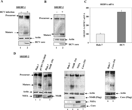
FIG. 1.
HCV induces proteolytic processing of SREBP-1 and -2. (A and B) Uninfected and HCV-infected Huh-7 cells were harvested at 48 h. Whole-cell lysates from Huh-7 and HCV-infected cells were fractionated by SDS-PAGE and immunoblotted with anti-SREBP-1/2 monoclonal antibodies. Lanes 1, Huh-7 cell lysates; lanes 2, HCV-infected cell lysates. Bottom panels represent the expression of HCV core protein as a marker of HCV infection, and actin served as an internal protein loading control. (C) Transcriptional stimulation of SREBP-1 mRNA in HCV-infected cells. Total cellular mRNA was analyzed by using SREBP-1-specific primers. Bar 1, Huh-7 cells; bar 2, HCV-infected cells. The values represent the means and standard deviations of two independent experiments performed in duplicate. (D) HCV proteins induce proteolytic processing of mature SREBP-2. Whole-cell lysates from Huh-7 cells (lanes 1, 4, and 8 and unnumbered lane between lanes 2 and 3) and cells expressing HCV nonstructural proteins, pCMV729-3010 (lane 2), HCV NS4B (1b) (lane 3), NS4B (3a) (lane 5), NS5A (lane 6), HCV core (1b) (lane 7), and core (3a) (lane 9) were fractionated by SDS-PAGE and immunoblotted with anti-SREBP-2 monoclonal antibodies. The bottom panel represents the expression of individual HCV marker proteins.

