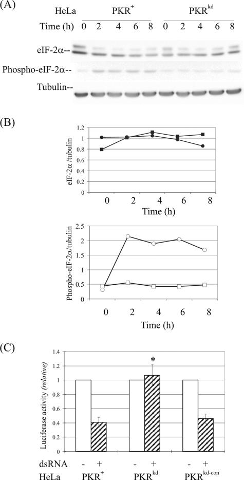
FIG. 4.
eIF-2α phosphorylation and protein expression following dsRNA treatment of PKR+ and PKRkd cells. (A) Western blot analysis. Whole-cell extracts were made at 0, 2, 4, 6, and 8 h after transfection with dsRNA, and 30 μg protein was analyzed in each lane. The membranes were probed with antibodies against eIF-2α or phospho-eIF-2α(Ser 51) and against α-tubulin as a loading control. (B) Quantification. Western blots were quantified by scanning densitometry. The upper panel shows the eIF-2α protein profile, and the lower panel shows the phosphorylated eIF-2α protein profile. Circles, HeLa PKR+; squares, HeLa PKRkd. (C) Protein expression. Luciferase reporter activity was measured in cells transfected in the absence (-) or presence (+) of dsRNA for 8 h, following transfection with pGL2-Control reporter DNA. *, P < 0.0001, Student's t test, PKRkd compared to PKR+ parent or PKRkd-con.

