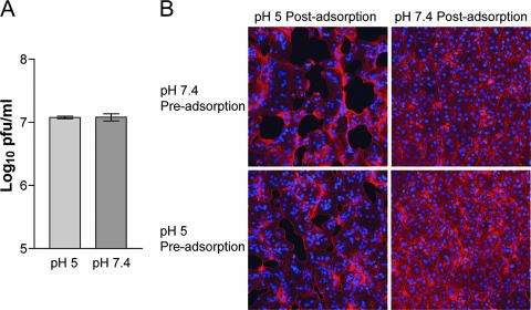
FIG. 1.
Effect of a brief low-pH treatment in the absence of target membranes on MV infectivity and syncytium formation. (A) Purified VACV WR MVs were exposed to pH 5 or pH 7.4 buffer for 3 min at 37°C, and the infectivity titer was determined by plaque assay. (B) Purified VACV WR MVs were exposed to pH 5 or pH 7.4 buffer for 3 min at 37°C (Pre-adsorption) and adsorbed to BS-C-1 monolayers for 1 h at 4°C. The cells were then immersed in either pH 5 or pH 7.4 buffer at 37°C (Post-adsorption). After 3 min, the buffer was replaced with EMEM containing cycloheximide and incubated for 3 h at 37°C. The cells were fixed and stained with Alexa Fluor 568-phalloidin and DAPI to display actin filaments and DNA, respectively.

