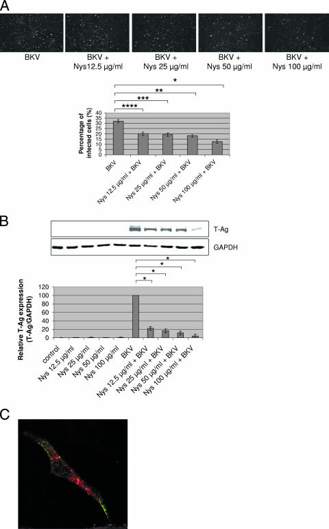
FIG. 4.
Nys interfered with BKV infection. HRPTEC were preincubated with Nys (12.5, 25, 50, and 100 μg/ml) for 1 h prior to coincubation with BKV (MOI, 0.5 FFU/cell) and Nys for 72 h. The medium was removed, and cells were washed three times with REBM with 0.5% FBS and incubated for another 48 h with fresh medium containing Nys. (A) After incubation, cells were fixed and analyzed by IF (magnification, ×10). T-Ag-positive cells were counted as BKV-infected cells, and the percentage was calculated against total cells. At least 500 cells were counted from three independent coverslips, and means and SE were calculated from two independent experiments. *, P < 0.01; **, P < 0.005; ***, P < 0.005; ****, P < 0.002. (B) After incubation, cells were harvested and analyzed by WB. Relative levels of T-Ag expression were detected by measuring the intensities with an Odyssey system and depicted as graph bars. The intensity of T-Ag expression was corrected by the intensity of GAPDH as the loading control. Means and SE were calculated from the results of two independent experiments. Control, HRPTEC were not incubated with either BKV or Nys; *, P < 0.001. (C) HRPTEC were preincubated with 100 μg/ml of Nys for 1 h prior to 8 h of coincubation with labeled BKV (MOI, 5 FFU/cell) and transferrin (10.0 μg/ml). After cells were fixed, Alexa Fluor 488-labeled BKV (green) and Alexa Fluor 633-conjugated transferrin (red) were observed by confocal microscope using a 63× lens objective.

