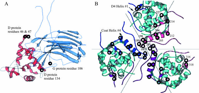
FIG. 5.
(A) Location of the second-site suppressor in the major spike and external scaffolding protein D1 subunit. Protein G and the D1 subunit are shown in light blue and red, respectively. The locations of the plaque formation mutations are depicted with circles and identified on the figure. (B) The clustering of the plaque formation mutation in the major coat protein at the threefold axis of symmetry. Coat proteins are depicted in blue, purple, and pink. The D4 external scaffolding protein subunit is depicted in cyan. Amino acid numbers are given on the pink coat protein subunit. Some of these second-site suppressors were previously isolated (17). Suppressors located at coat protein residues 12, 81, 138, 333, and 426 are not depicted.

