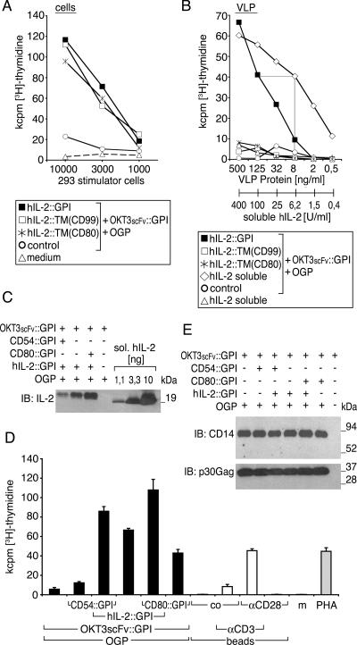
FIG. 5.
VLP expressing GPI-anchored hIL-2 modulate human T-lymphocyte activation. The biological activity of IL-2 fusion proteins on producer cells (A) and resulting VLP (B) was determined upon cotransfection with OKT3scFv::GPI. As controls, control vector-transfected producer cells and particles thereof, OKT3scFv::GPI-decorated particles supplemented with soluble hIL-2, soluble hIL-2 alone, or medium alone was used. PBMC (105) were incubated with titrated amounts of the indicated 293 cells starting with a cell number of 1 × 10⁴ in A or VLP or VLP/soluble hIL-2 mixtures starting with 500 μg/ml of particles in B. Cell proliferation was determined after 4 days by [3H]thymidine uptake. Data show mean values of triplicate cultures representative for two independently performed experiments. (C) A total of 360 ng of VLP coexpressing hIL-2::GPI (same particles as in B, D, and E) was compared to a titration curve of soluble hIL-2 (1.1, 3.3, and 10 ng) by immunoblotting (IB) using the hIL-2-specific mAb IL-2.1F10. Binding of antibodies in C was visualized by HRP-conjugated secondary reagents, followed by a luminol-based detection reaction (Perkin-Elmer, Boston, MA). Molecular mass is indicated in kilodaltons. One out of several typical experiments is shown. (D) Human PBMC (105) were cocultured with supernatants of stably OKT3scFv::GPI-expressing 293 cells that were cotransfected with original MoMLV gag-pol (OGP), CD80::GPI, CD54::GPI, and hIL-2::GPI or control vector combined as indicated (black bars). Microbeads (105/well) substituted with CD3 mAb, CD28 mAb, control mAb (co), or combinations thereof were used for comparison (white bars). PHA or culture medium (m) served as additional controls (gray bars). Cell proliferation was determined after 4 days by [3H]thymidine uptake. Data show mean values ± standard deviations of triplicate cultures representative of several independently performed experiments. (E) The amounts of VLP in the various supernatants used for proliferation assays in A were determined by immunoblotting for OKT3scFv::GPI using a CD14 mAb and for viral core proteins using a p30 Gag mAb. Binding of antibodies was visualized by HRP-conjugated secondary reagents, followed by a luminol-based detection reaction (Perkin-Elmer). Molecular mass is indicated in kilodaltons.

