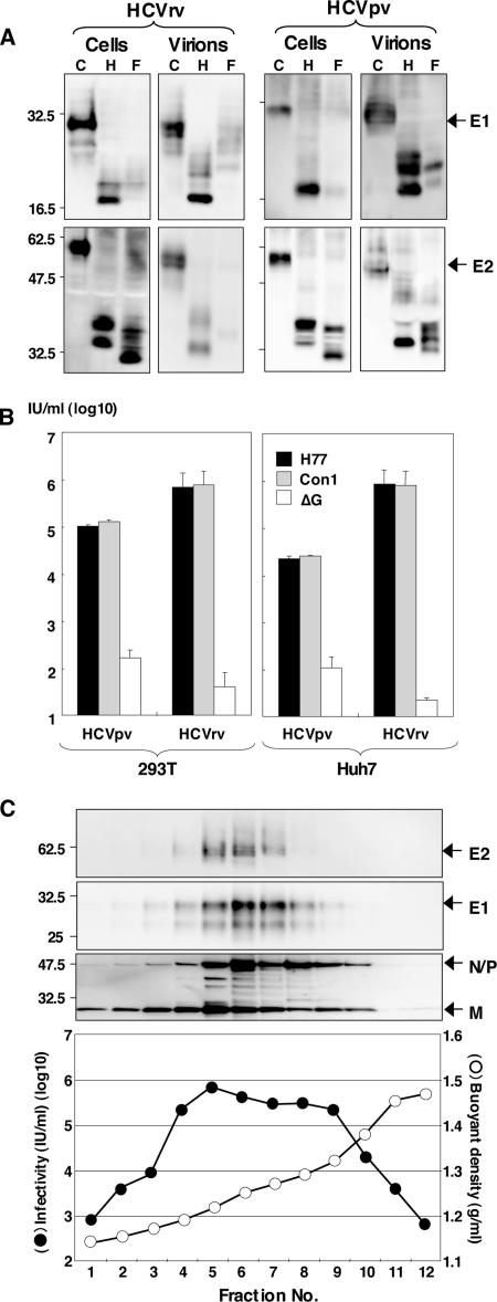
FIG. 3.
Characterization of HCVrv and HCVpv. (A) The E1 and E2 proteins of the H77 strain expressed in 293T cells and incorporated into the particles of HCVrv and HCVpv were either untreated (C) or treated with endoglycosidase H (H) or peptide-N-glycosidase F (F). Following fractionation on sodium dodecyl sulfate-polyacrylamide gel gels, the glycoproteins were detected by immunoblotting with anti-E1 (BDI198) and anti-E2 (AP33) monoclonal antibodies. (B) The infectivities of HCVrv and HCVpv bearing HCV envelope proteins of genotypes 1a (H77 strain) and 1b (Con1 strain) generated in 293T or Huh7 cells were determined with Huh7 cells. The envelope-less VSV (ΔG) was used as a control. (C) (Top) CsCl gradient sedimentation of HCVrv produced in 293T cells. The supernatant was fractionated from the top of the gradient and analyzed by immunoblotting with anti-E2, anti-E1, and anti-VSV antibodies. (Bottom) The infectivity (filled circles) of each fraction was determined after the removal of CsCl with column purification. Fraction densities (open circles) are expressed in grams/milliliter.

