FIG. 8.
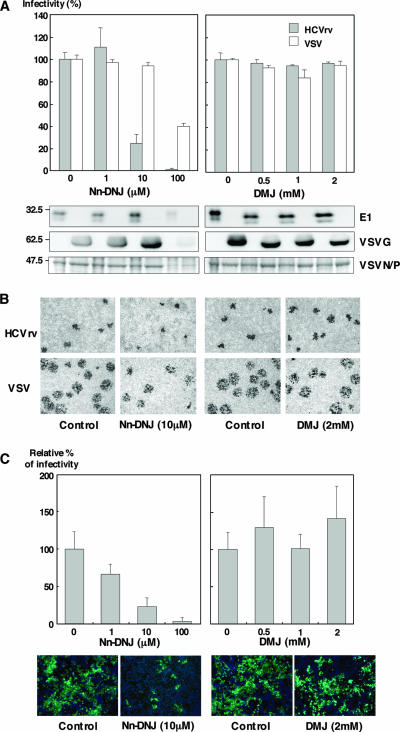
Effects of ER α-glucosidase inhibitors on the infection with HCVrv and HCVcc. (A) (Top) Production of HCVrv and VSV in the presence of Nn-DNJ (left) or DMJ (right). Huh7 cells infected with HCVrv and VSV at MOIs of 0.1 and 0.01, respectively, were treated with various concentrations of Nn-DNJ or DMJ. Seventy-two hours (HCVrv) or 24 h (VSV) postinfection, culture supernatants were collected and titrated on Huh7 cells by a focus-forming assay. The results shown are from three independent assays, with the error bars representing the standard deviations. (Bottom) Purified viruses generated in Huh7 cells treated with Nn-DNJ or DMJ were analyzed by immunoblotting with anti-E1 (BDI198) and anti-VSVG (ab34774) or Coomassie staining. (B) Focus formation of HCVrv and VSV in the presence of Nn-DNJ or DMJ. Huh7 cells were infected with HCVrv or VSV, treated with Nn-DNJ (10 μM) or DMJ (2 mM) prior to an overlay of culture media containing 0.8% of methylcellulose, and stained with an anti-VSV N antibody after fixation at 72 h (HCVrv) and 24 h (VSV) postinfection. (C) (Top) Production of HCVcc in the presence of Nn-DNJ (left) or DMJ (right). Huh7.5.1 cells infected with HCVcc at an MOI of 0.01 were treated with various concentrations of Nn-DNJ or DMJ. Culture supernatants were collected and titrated by a quantitative core enzyme-linked immunosorbent assay at 96 h postinfection. (Bottom) Immunofluorescence assay of HCVcc infection in the presence of Nn-DNJ or DMJ. Huh7.5.1 cells were infected with HCVcc atan MOI of 0.01, treated with 10 μM of Nn-DNJ or 2 mM of DMJ prior to an overlay of culture media containing 0.8% of methylcellulose, and stained with an anti-NS5A antibody and Alexa 488-conjugated secondary antibody after fixation at 96 h postinfection. Cell nuclei were stained by Hoechst 33258. Pictures were taken using a fluorescence microscope by double exposure of the same fields with filters for Alexa 488 or Hoechst 33258.

