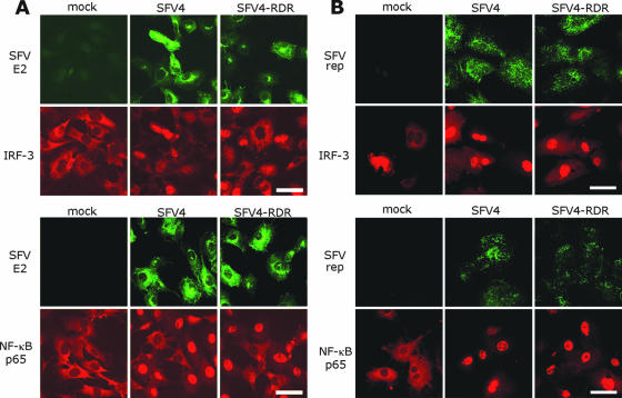
FIG. 5.
B6 MEFs were infected at a MOI of 1 (A) or 50 (B) with SFV4 or SFV4-RDR or were mock infected. Cells were fixed at 5 hpi (A) or 3 hpi (B) and immunostained with antibodies to SFV E2 or replicase (green fluorescence) and either IRF-3 (upper panels, red fluorescence) or NF-κB p65 (lower panels, red fluorescence). Bar, 50 μm.

