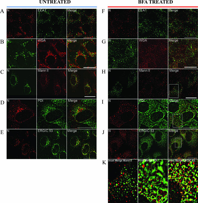
FIG. 2.
Colocalization of HTNV N with ERGIC-53 and redistribution of N with BFA. Vero E6 cells were infected with HTNV at an MOI of 0.1, and after 3 days slides were acetone fixed (except for Mann II staining, in which case paraformaldehyde was used). Prior to fixation, slides F to K were treated with BFA for 1 h as described in Materials and Methods. Slides were costained with WGA (B and G) or antibodies (anti-N monoclonal E-314 [green] or polyclonal no. 143 antibody [red]) against EEA1 (A and F), Mann II (C and H), PDI (D and I), or ERGIC-53 (E and J) as described in Materials and Methods. Enlarged merged images of the insets in panels H, I, and J are presented in panel K. Scale bars, 20 μm, using 100× objectives (Leica confocal microscope).

