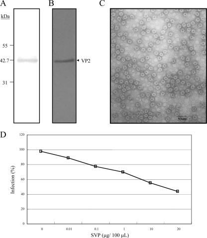
FIG. 2.
Purity analysis of VP2-441 SVP prepared by IMAC and inhibition of IBDV infection of DF-1 cells by SVP. IMAC-purified SVP was concentrated through a centrifugal filter (100 kDa) and analyzed by SDS-PAGE with silver staining (A) and Western blotting with anti-VP2 polyclonal antibody (B). Molecular mass standards are shown on the left in kDa. (C) Electron micrograph with ×100,000 magnification (scale bar, 50 nm) and negatively stained with 2% uranyl acetate. (D) Dose-dependent inhibition of IBDV infection by SVP. DF-1 cells were preincubated with different concentrations of SVP (0, 0.01, 0.1, 1, 10, and 20 μg/100 μl) for 1 h at 4°C and then infected with 10 TCID50 of IBDV P3009 for 1 h. CPE was determined using crystal violet staining 96 h after infection. Two separate experiments showed similar results.

