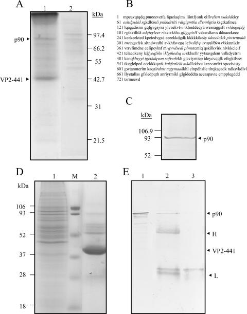
FIG. 5.
Isolation of cHsp90 with affinity for SVP. (A) Affinity isolation of a p90 protein with SVP as the IMAC ligand. A procedure described in the text was set up for affinity isolation of putative IBDV receptors using SVP as the ligand bound to the immobilized Ni2+ ions. In brief, total proteins from DF-1 cells were passed through the affinity chromatographic column. After washing with two wash buffers, the elution of SVP and its associated factors was accomplished by using an IMAC elution buffer. The fractions collected were concentrated, and 40 μl of the concentrate (lane1) and, as a control, 50 μl of the concentrated elution fraction obtained in the absence of SVP (lane 2) were separated by SDS-12.5% PAGE and stained with Coomassie blue (left). Positions of a p90 protein and the monomer VP2-441 that forms SVP are indicated on the left. (B) Identification of p90 by mass spectrometry. The protein sequence of cHsp90 was derived from Protein Data Bank accession number P11501. Peptide sequences of p90 identified by mass spectrometry are shown in italics. (C) Identification of p90 as cHsp90 by Western blotting. An aliquot of 40 μl of the concentrated elution from the affinity column chromatography with SVP was separated by SDS-12.5% PAGE, transferred onto a PVDF membrane, and incubated with an anti-Hsp90 MAb. The membranes were then incubated with a second antibody coupled to alkaline phosphatase and developed in a buffer containing NBT and BCIP. Molecular mass markers are indicated on the left. An arrow indicates the position of p90 (cHsp90). (D) Isolation of p90 from DF-1 cells by immunoprecipitation. Total proteins from DF-1 cells (lane 1) and the precipitated immune complexes (lane 2) were separated by SDS-12.5% PAGE and stained with Coomassie blue (left). The precipitated immune complexes in the presence of SVP (lane 2) and without SVP (lane 3) were separated by SDS-12.5% PAGE, transferred onto a PVDF membrane, and incubated with anti-Hsp90. (E) Western blots were developed as described above. As expected, cHsp90 (p90) was identified in the total DF-1 lysate (lane 1). Molecular mass markers are indicated on the left. Arrows indicate positions of proteins of the immune complexes.

