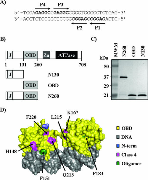
FIG. 1.
Sequence of site II and purified proteins used in this study. (A) Nucleotide sequence of site II located in the SV40 origin of replication. The positions of the four pentanucleotide binding sites, P1 to P4, are indicated. (B) Schematic representation of the 708-aa-long SV40 large T-ag and subfragments thereof used in this study. The following functional domains are shown: J-domain (J; aa 1 to 82), OBD (aa 131 to 260), zinc finger (Zn; aa 302 to 320), and ATPase/helicase domain (aa 418 to 616). (C) Coomassie-stained 15% SDS-PAGE gel of the purified T-ag fragments used in this study. Three-microgram portions of each protein, N260, OBD, and N130, were analyzed. (D) Structure of two OBD molecules bound independently to an oligonucleotide containing P1 and P3 (PDB, 2NTC) (37). Given that P1 and P3 are in opposite orientations, both faces of the OBD can be seen in this structure. Amino acids changed in class 4 mutants (pink) or located at a putative OBD oligomerization interface (green) (37) are indicated. The N-terminal residue of the OBD where the N-terminal domain would be connected is colored in blue.

