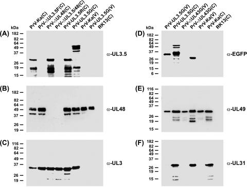
FIG. 2.
Western blot analyses. RK13 cells were harvested 16 h after infection with the indicated viruses (MOI = 5). Lysates of infected and noninfected cells (C) and of sucrose gradient-purified virions (V) were separated by SDS-PAGE. Blots were incubated with monospecific rabbit antisera against the C-terminal part of the UL3.5 gene product (A), the viral tegument components encoded by UL48 (B) and UL49 (E), the nonstructural UL3 (C) and UL31 (F) proteins, or EGFP (D). Molecular mass markers are indicated at the left.

