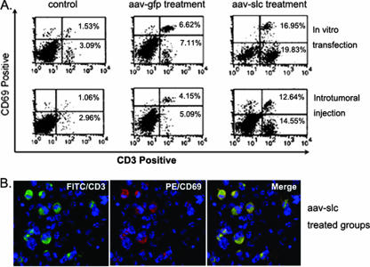
FIG. 5.
The infiltrated T cells are mostly activated cells. In order to determine the bioactivity of the infiltrated T cells, the collected cells were also double stained with FITC-anti-CD3 and PE-anti-CD69. Isotype-matched antibodies were used as a control. In comparison with the control and rAAV-GFP treatment groups, significantly higher numbers of CD3+ CD69+ T cells were found in the infiltrate of SLC-treated mice by flow cytometric evaluation. Most CD3-positive T cells (green) are CD69 positive (red) in the rAAV-SLC-treated group and displayed double staining (yellow).

